
Laser capture microdissection is used to excise cells or cell subsets from sample tissues. The resulting dissections can be analyzed by multi-omic applications downstream.
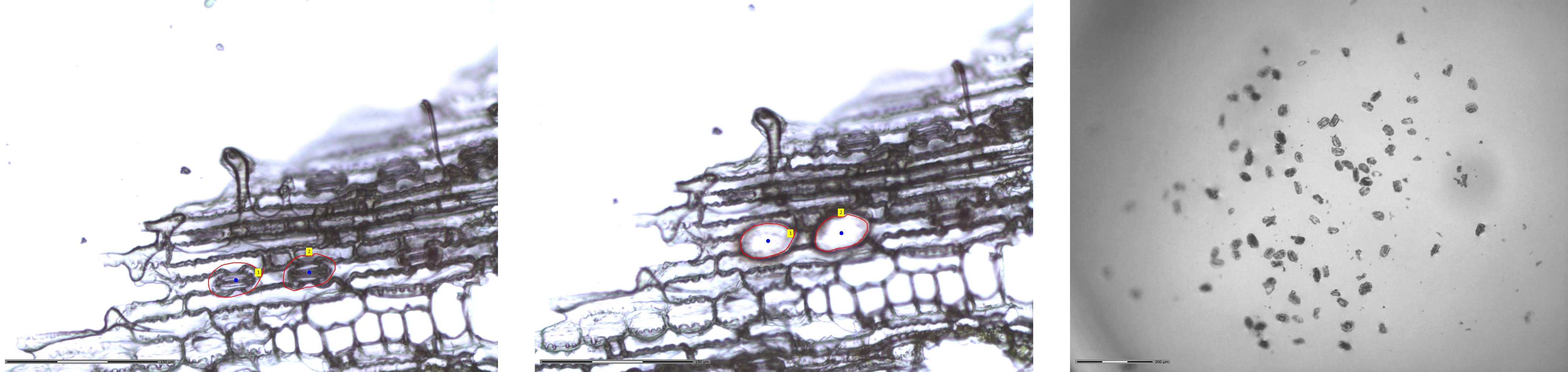
1. Sample Preparation
- Isolate spatially resolved samples from multicellular tissues or biofilms
- Cryosection or create a micrograph
- Fix sample cross-sections with ethanol and then dehydrate
2. Cryotomy
- Embed sample in an appropriate material (optimal cutting temperature, hydroxypropyl-methyl cellulose polyvinylpyrrolidone, etc.)
- Prepare thin sections with a cryostat
- Transfer thin section(s) to a polyethylene naphthalate membrane slide
3. Laser-Microscope Dissection
-
Scan slide and align with collection device
-
Image for target identification
-
Dissect subsections and collect voxels
-
RNA – short collection
-
Protein – longer collection
-
Collect images
4. Further Analyses
Contact
Will Chrisler, william.chrisler@pnnl.gov
Fluorescence-Activated Cell Sorting (FACS) is used to characterize cell populations directly or prepare sample subsets for downstream analysis.
1. Sample Preparation
- Determine if live or fixed cells are required
- Apply stain or dye:
- immunostaining
- vitality stains
- organelle stains
- fluorescent proteins
- activity-based probes
- Dilute and declump to generate a single-cell suspension
- Remove cell wall (if required)
2. Initial Cell Population Analysis
- Analyze population by cell size/complexity
- Sort subset on gate criteria
- Verify expectation/accuracy of sorting by confocal microscopy
3. Sorting
- Isolate single cells into 96/384-well formats
- Collect bulk subpopulation(s)
- Quantify abundance or proportions as an endpoint
4. Further Analyses
- Mass spectrometry proteomics
- Transcriptomics
- Nanodroplet processing in one pot for trace samples
- Nanoscale secondary ion mass spectrometry
- Fluorescence in situ hybridization
- Activity-based probes
Contact
Will Chrisler, william.chrisler@pnnl.gov
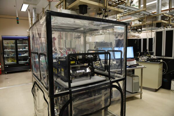
cellenONE is high-precision single-cell isolation and dispensing system that enables the accurate isolation of individual cells from heterogeneous samples for further analysis. The system operates at a lower pressure relative to that for FACS for handling delicate samples. The workflow involves the precise sorting of single cells and controlled liquid handling down to a picoliter size, all with piezoelectric dispensing for very small volumes. cellenONE is a robust tool for applications in genomics, transcriptomics, and proteomics to aid our understanding of cell heterogeneity and complex biological processes.
1. Sample Preparation
- Determine if live or fixed cells are required
- Apply stains or dyes:
- immunostaining
- vitality stains
- organelle stains
- fluorescent proteins
- activity-based probes
- Dilute and declump to generate a single-cell suspension
- Remove cell wall (if necessary)
2. Quality Check
- Analyze population by cell size/signal
- Sort a subset on gate criteria
- Collect images (of each sorted cell or item)
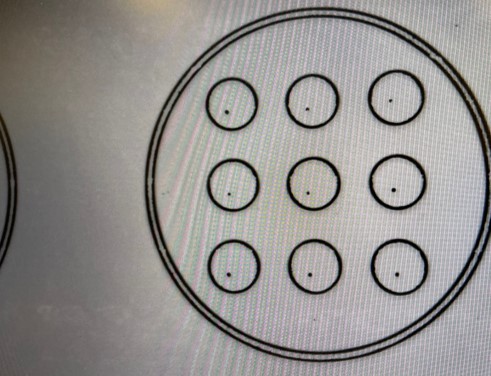
3. Sorting and picoliter dispensing Applications
- Sorting single cell into a 96- or 384-well format on different type of modified glass slides like nanoPOTS chip or indium tin oxide-coated glass slide.
- Dispensing picoliter of samples or reagents onto amino functional glass slide or Cryo-EM Grids.
4. FURTHER ANALYSES
- mass spectrometry
- transcriptomics
- Nanodroplet Processing in One Pot for Trace Samples (nanoPOTS)
- Nanoscale secondary ion mass spectrometry
- Fluorescent in situ hybridization