

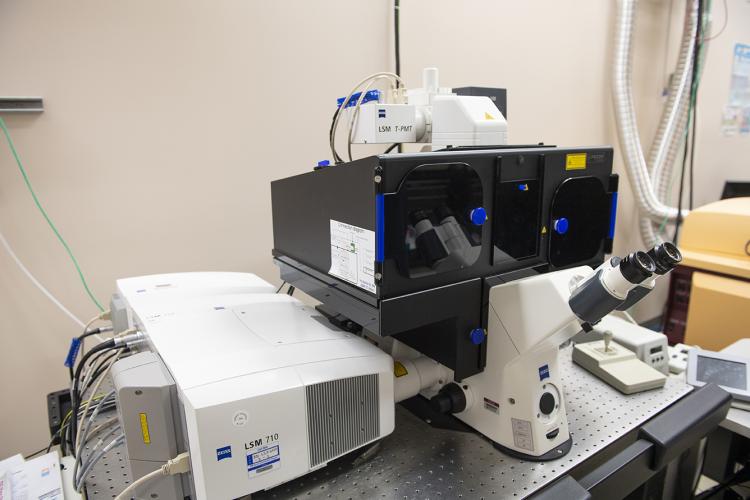
Super resolution structured illumination and Airyscan fluorescence microscope
The Airyscan module, mounted on the confocal fluorescence microscope, is an addition to EMSL’s structured illumination microscopy system. It provides 3D imaging with a superior detection sensitivity and speed compared when compared to the standalone structured illumination microscopy system or classic confocal microscope. The enhanced imaging allows researchers to image live or intact hydrated cells in 3D in their near native form, which is critical for understanding cellular systems. All commonly used fluorescent proteins or dyes can be used for imaging by this system. It can detect weak fluorescent signals, such as those emitted by fluorescent proteins expressed at low levels or by chlorophyll and capture dynamic cellular processes in real-time with 150 nanometer lateral resolution. Studies that use this system focus on the spatial and temporal gene expression patterns, using fluorescence in situ hybridization and targeting enzymes responsible for wood degradation by fungal hyphae or nitrogenases in endophytic bacteria, as well as quantifying lipid production in microorganisms.
Research application
- Supporting the Cell Signaling and Communication Integrated Research Platform, the system supports studies investigating the spatial and temporal dynamics of proteins and other molecules in individual plant cells within intact tissues and helps determine the functional significance of microbial community structures. Airyscan is also used to study spatial and temporal protein expression patterns, using fluorescent proteins or antibodies, targeting proteins critical for stress response in plants or enzymes that are important for cellulose degradation in microorganisms.
Available resources
- Stochastic Optical Reconstruction Microscopy / Photoactivated Localization Microscopy - Super Resolution Fluorescence Microscope
- Multiphoton Confocal Microscope
- Lattice Light Sheet Microscope
Tips for success
- The system can image a wide variety of samples. The sample specimen may be on microscope slides, coverslips, in cell culture dishes under growth medium, or on dry surfaces.
- A selection of microscope objectives are available for imaging the different samples, such as air, water immersion, and oil immersion with high and low magnifications.