

TerraForms: Pore-Scale Micromodels
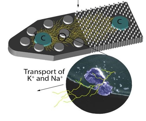
The Environmental Molecular Sciences Laboratory’s (EMSL’s) pore-scale micromodels simulate soil porosity and aggregate size distribution on a microfluidic platform. These TerraForms are suitable for microbial (bacterial and fungal) incubations for investigating pore-scale effects on resulting growth. The micromodels have been used to study anaerobic hotspot and hot moment formation by microbial growth, fungal hyphae development, and the effects of carbon sources on extracellular polysaccharide-induced water retention within soil pores and to investigate microbial potential for biomining critical minerals for harvesting rare earth elements.
Mineral-amended micromodels can be created to simulate a specific soil mineralogy, including specific critical minerals. These micromodels have been used to investigate fungal mineral weathering, the distribution of microbial exudates around a carbon hotspot, and the changes in mineral chemistry resulting from biotic mineral weathering.
Research application
Supporting the Biogeochemical Transformations Integrated Research Platform, these resources help unlock how nutrients, critical minerals, and materials move and change in the environment. EMSL’s microfluidic technologies allow scientists to create synthetic soil habitats to understand and to study microbe–mineral interactions for investigating soil biogeochemical inorganic nutrient cycling and biomining critical mineral elements.
Supporting the Terrestrial-Atmospheric Processes Integrated Research Platform, TerraForms enables belowground and aboveground volatile organic compounds to be identified for your science.
Supporting the Biomolecular Pathways Integrated Research Platform, TerraForms pore-scale micromodels enable the detection of gases and volatile organic compounds using the real-time mass spectrometry.
Available resources
- A HORIBA Jobin Yvon Raman spectroscopy system with 532 and 632 nm lasers combined with a Nikon Eclipse Ti epifluorescence microscope for mineral and microbe characterization and imaging.
- A stand-alone Nikon Eclipse Ti epifluorescence microscope for mineral and microbe characterization and imaging.
- A Nikon AZ100 multipurpose zoom fluorescent microscope connected to a motorized stage and charge-coupled device (CCD) camera for imaging larger samples at 0.8–20 μm resolution.
- A Class 1000 clean room microfabrication facility with equipment (e.g., mask aligner, plasma dry etch, anodic bonding, etc.) that allows the fabrication of microfluidic pore structures in silicon, PDMS (polydimethylsiloxane), glass, etc.
Tips for success
All pore-scale micromodels are compatible with mass spectrometry imaging such as matrix-assisted laser desorption ionization mass spectrometry imaging, nanospray desorption electrospray ionization, secondary ion mass spectrometry (SIMS), nanoSIMS, and scanning electron microscopy. Request pore-scale micromodels to investigate spatial metabolites and the carbon distribution resulting from microbial growth or to investigate hotspots and hot moments within a soil-like environment.
Contributing teams and resources
EMSL develops and deploys capabilities for the user program by conducting original research independently or in partnership with others and by adapting/advancing science and technologies developed outside EMSL. In some instances, EMSL directly deploys mature capabilities developed by others where there is value for the EMSL user community. The following grants/activities, principal investigators, and teams contributed to the development of this capability:
- Arunima Bhattacharjee, DE-AC05-76RL01830, Environmental Molecular Sciences Laboratory
- Kirsten Hofmockel, FWP 70880, U.S. Department of Energy, Office of Science, Biological and Environmental Research Program.
Related publications
- Bhattacharjee, A., et al. 2022. "A Mineral-Doped Micromodel Platform Demonstrates Fungal Bridging of Carbon Hot Spots and Hyphal Transport of Mineral-Derived Nutrients." Mycology 7 (6): e00913-22. https://doi.org/10.1128/msystems.00913-22.
- Bhattacharjee, A., et al. 2023. "Fungal Organic Acid Uptake of Mineral-Derived K Is Dependent on Distance from Carbon Hotspot." Mycology 14 (5): e00956-23. https://doi.org/10.1128/mbio.00956-23.
- Lukowski, J., et al. 2021. "Expanding Molecular Coverage in Mass Spectrometry Imaging of Microbial Systems Using Metal-Assisted Laser Desorption/Ionization." Microbiology Spectrum 9 (1). https://doi.org/10.1128/spectrum.00520-21.
- Guo, Y.-S., et al. 2024. "Accessing Fungal Contributions to the Birch Effect: Real-Time Respiration from Pore-Scale Microfluidics." Microorganisms 12 (11): 2295. https://doi.org/10.3390/microorganisms12112295.